We got the package! The cow eye came in the the mail from Home Science Tools.

To wrap up our human eye unit in school, we decided to get a hands on experience by handling and dissecting a real eye. The kit came complete with a scalpel, scissors, dissection guide, a dissecting tray, and, of course, the cow eye.
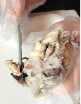
First, we opened the eye package and placed the eyeball on the dissecting tray to examine it. We located the optic nerve, the cornea, and the sclera.
First, we opened the eye package and placed the eyeball on the dissecting tray to examine it. We located the optic nerve, the cornea, and the sclera.
 |  |
We then cut away the excess fat and muscle using the scalpel and scissors. We wore gloves to protect our skin from the formaldehyde that the eye was preserved in.
 |  |
It was so much fun using a scalpel!
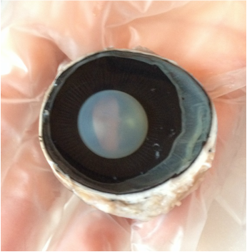
Once all the fat and muscle were removed, we cut the eye in half using the scissors and looked at the vitreous humor inside. We received a well preserved eye, so the vitreous humor was still clear and gelatinous.The anterior of the eye contains the cornea, lens, iris, and ciliary body. The posterior of the eye contains the retina and optic nerve.

We removed the vitreous humor carefully so it did not remove the retina (the white film in the bottom half of the eye shown above) with it. Then we took the anterior part of the eye, and using the scissors, we cut the cornea out of the eye.
 | 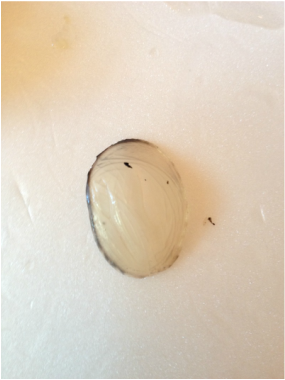 |
The cornea is tough and thick. It protects the eye. With the cornea removed, we could easily see the iris and the lens. The iris opens wide when it is dark and becomes small when it is bright.
We turned the anterior half over and as we removed the lens we could clearly see the ciliary body, which allows the lens to change shape as it focuses on different objects close and far. We were able to place the lens over a piece of paper with words on it to see how the lens works by magnifying objects.
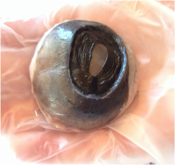
Next, we looked at the posterior half of the eye. The retina covers the sclera and contains photo receptors that collect light and turn it into electric pulses that travel to the optic nerve and into the brain. There are no photo receptors where the optic nerve attaches to the eye creating a blind spot. The retina is connected to the eye only where the optic nerve comes into the eye.

The choroid coat is very colorful and shiny in a cow eye. This allows light to reflect back onto the retina at night, allowing them to see better when it is dark. This is responsible for the "glowing eyes" of animals at night. Human eyes have a dark colored choroid coat, minimizing the reflection of light back onto the retina. The extra light on an animal's retina causes distorted images.
This dissection project was very awesome! We look forward to our next dissecting project!!!
 RSS Feed
