We got the brain! We got to dissect it. Here's what we learned.

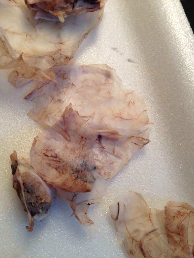
After we put on our surgical gloves, we took the sheep brain out of the package and laid it on our styrofoam tray. The brain had been preserved in formaldehyde so it was a little stinky. We first noticed that the brain was covered with a thin white translucent membrane. It looked much like the skin that peels after a bad sunburn. We thought it would be easy to peel off. This meninges has three layers, but we could only see the outermost layer, the dura mater. This membrane supports and protects the brain.

We soon found out that it was tougher than us so we had to get our dissecting scissors out. Finally after careful cutting it came off. We noticed that the dura mater is full of blood vessels.
 |  |
Having removed the dura mater we could see the brain a lot more clearly.
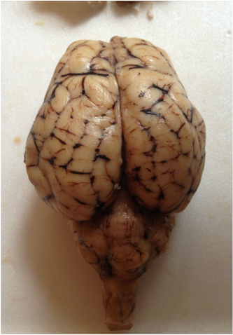
We were able to see the frontal lobe (the front of the brain), temporal lobe (at the temple right above the ears), parietal lobe (at the top back of the head), and occipital lobe (at the base of the head). These lobes form the cerebrum or cortex of the brain. The cerebrum is where information is stored and it oversees many everyday functions. We could also easily see where the two hemispheres were divided, called the longitudinal fissure. We looked at the cerebellum (right below the occipital lobe sitting on the spinal cord). The cerebellum controls movement, balance, and contains almost half of the brain's neurons. Damage to this area may cause lack of balance or slow movement.
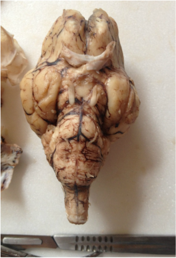
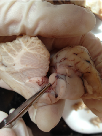
On the underside of the brain we saw the olfactory bulbs responsible for sensing odors.
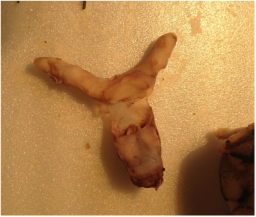
We located the optic chiasm, which is where the optic nerves from the eyes come together and cross in an "X" shape.
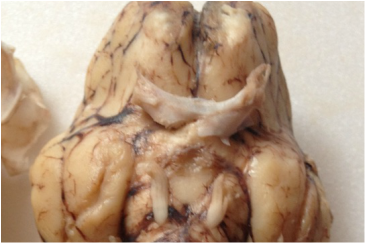 |  |
The infundibulum is the white area right below the optic chiasm where the pituitary gland is attached prior to removing the dura mater. The pituitary gland controls growth, body temperature, reproduction, and thyroid functions, among other things.
Right next to the infundibulum are the oculomotor nerves. These control the eyeball and eyelid movement.
The pons are below the oculomotor nerves. The pons control many functions such as hearing, taste, balance, sensation of touch and pain, as well as motor functions like chewing and swallowing.
The hypothalamus is next to the pons, about the area that the oculomotor nerves enter the brain, and it controls hunger, thirst, sleep, attachment and parenting behaviors, and body temperature.

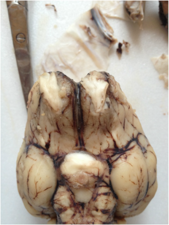
When we pulled the cerebellum back from the cortex, it exposed the colliculi. There is still a lot unknown about this organ, but they believe it helps with eye movement and processing hearing.
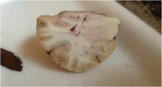
It was time to cut the brain in half. The left and right hemispheres of the brain are basically separate and can practically be pulled apart until the cerebellum and spinal cord but the complete underside needed to be cut.

Once the brain was cut in half, we could clearly see corpus callosum, which connects the left side of the brain to the right. It is the white horizontal tissue with a slight curl at the end.
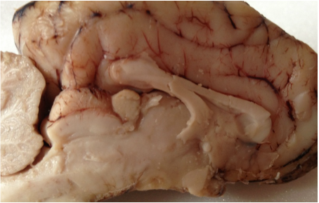 |  |
We found the pineal gland (the round shaped tissue located right under the corpus callosum), which produces melatonin in order to regulate sleep cycles. Remember how the hare's hair changes color according to the amount of light that its eyes receive? Our melatonin production is also based off of the amount of light that the photo sensors in our eyes receive. We make just enough melatonin to stay awake during the day, and stay asleep at night.
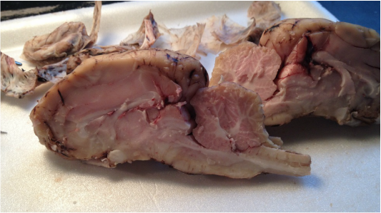
The last thing we did was cut the brain's cortex in half, perpendicular to the first cut. This allowed us to more clearly see the white and gray matter of the brain.
Gray matter (the darker tissue) allows us to think, show self control, compute and make decisions, have memories, emotions - all muscle control and sensory input. White matter allows communication within the gray matter. It is white due to the coating on the nerve endings, myelin.
The brain is so amazing! All those functions stuffed into one organ - a mega-computer!
 RSS Feed
